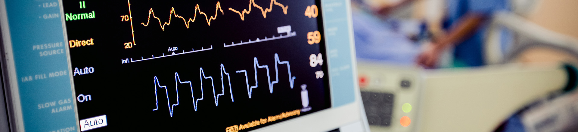
Cardiovascular Diagnostics & Testing
MarinHealth provides state-of-the-art cardiovascular testing, imaging, and diagnostics:
- To diagnose an urgent condition, such as an apparent heart attack
- To screen people who have been experiencing chest pain or shortness of breath or have an intermediate to high risk for heart disease
- To proactively detect early signs of heart disease
Diagnostic tests to detect heart disease are classified as either noninvasive or invasive. Noninvasive tests do not require penetration of the skin or a body cavity beyond the use of an injection. Invasive tests involve puncturing the skin and entering major blood vessels, and must be performed at MarinHealth's Haynes Heart & Vascular Institute at MarinHealth Medical Center, usually on an outpatient basis.
View a list of cardiovascular tests available at each of our locations, or learn more about our Cardiovascular Performance Center.
Ambulatory Heart Monitors
This type of testing is used for patients who experience transient symptoms
that suggest a cardiac arrhythmia. Current methods of rhythm monitoring
include zio patch XT monitoring or zio patch AT monitoring which is a
mobile cardiac telemetry monitor. These are typically worn for 14 days,
and a comprehensive report is available to the provider.
Calcium Scoring
This is a noninvasive way of obtaining information about the location and
extent of calcified plaque deposits, a marker for heart disease, in the
coronary arteries.
Cardiac MRI
Cardiac magnetic resonance imaging (MRI) is a cutting-edge, noninvasive
diagnostic tool that provides high-resolution images of the heart while
beating. This allows for a thorough and incomparable evaluation of myocardial
structure, tissue composition, and function. Our heart specialists and
MRI team use radio waves within the MRI scanner to create highly detailed
still and dynamic digital renderings of your heart.
Computed Tomography Angiography (CTA)
This imaging technique combines rotating X-ray equipment with a digital
computer to produce remarkably clear, detailed, cross-sectional imaging
of the heart and coronary arteries. With the images produced by the CTA
we are currently able to collaborate with Heartflow® to produce a 3D structure of the coronary arteries.
Coronary Angiography
This technique uses X-rays and intravenous contrast to visualize the circulatory
system throughout the body, including the heart.
Coronary Reactivity Test
This is a type of angiography procedure designed to examine the blood vessels
in the heart and how they respond to different medications. This type
of test helps a physician to understand blood vessel reactivity dysfunction.
Echocardiogram
An echocardiogram–often called “echo” or cardiac ultrasound,
provides a graphic outline of the heart’s structures, movement,
and function and helps with the diagnosis of arrhythmias.
Electrocardiogram (EKG)
An electrocardiogram provides physicians with important information about
the heart’s rhythm by recording its electrical signals. Electrodes
are placed in various places on the patient’s chest and limbs. It
is a non-invasive, painless, and safe diagnostic tool in electrophysiology.
Electrophysiology Study
This is a minimally invasive procedure that involves placing specialized
catheters in the heart via a patient’s blood vessels. The specialized
catheter allow a physician to see the electrical conduction of the heart
with greater detail that the EKG can provide. During the study, a physician
can provoke an arrhythmia using several methods. An EP study can provide
a definitive diagnosis of an arrhythmia and information essential in the
selection of the appropriate treatment.
Exercise/Pharmacologic Stress Tests
Several types of stress tests can be conducted depending on your overall
health and mobility. Exercise stress tests involve exercising on a treadmill
or stationary bicycle. Pharmacologic stress tests involve the injection
of a medication that mimics the effect of exercise on the heart. Regardless
of whether exercise or medications are used to speed the heart rate, we
can evaluate function in a variety of ways.
Intravascular Ultrasound Tests (IVUS)
Usually performed non-invasively, vascular tests detect the presence, severity,
and general location of vascular and arterial disease.
Myocardial Perfusion
A myocardial perfusion test evaluates the blood flow through the coronary
arteries to the heart muscle using a radioactive tracer. The completed
exam consists of intravenous access, resting images, a stress test, and
stress imaging.
Peripheral Vascular Angiography
This is done to detect narrowing or blockages in the blood vessels caused
by peripheral artery disease (PAD).
Stress Echocardiogram
A stress echocardiogram combines the ECG and heart function. It is an imaging
modality that shows the heart in action to measure and evaluate both the
electrical activity and the function and strength of the muscle under stress.
Stress Electrocardiogram (Stress ECG/Treadmill)
An ECG allows your physician to compare your heart’s electrical activity
at rest and under physical exertion. Stress ECGs are often combined with
imaging techniques.
Dobutamine Stress Echocardiography
This is a testing method used to evaluate blood flow in the coronary arteries
by administering dobutamine.
Transesophageal Echocardiogram
This is an alternative way to perform an echocardiogram. A specialized
probe containing an ultrasound transducer at its tip is passed into the
patient’s esophagus to assess how well the heart is working.


.jpg)
.jpg)
-west-wing_Option-1.jpg)
-south-wing_Option-1.jpg)


/
View All
Testing & Imaging Practitioners
Testing & Imaging Locations
-
 MarinHealth Cardiovascular Medicine | A UCSF Health Clinic
2 Bon Air Road
More Information
Suite 100
Larkspur, CA 94939
415-927-0666 -
MarinHealth Cardiovascular Medicine | A UCSF Health Clinic
75 Rowland Way
More Information
Suite 250
Novato, CA 94945
415-878-2910 -
MarinHealth Cardiovascular Medicine | A UCSF Health Clinic
651 First Street West
More Information
Suite L
Sonoma, CA 95476
707-935-1470 -
MarinHealth Cardiovascular Medicine | A UCSF Health Clinic
335 South McDowell Boulevard
More Information
Petaluma, CA 94954
707-293-1110