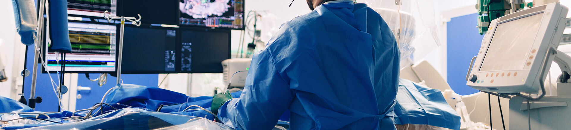
Electrophysiology Procedures
Choosing whether to manage a rhythm disorder with medications or procedures is a personal decision, as well as a medical one. Our specialists at MarinHealth's Haynes Heart & Vascular Institute will consult with you to explain the pros and cons of each approach and help guide you through the decision. If and when you are scheduled for a procedure, your physician will provide you with detailed information on what to expect.
Electrophysiology Study
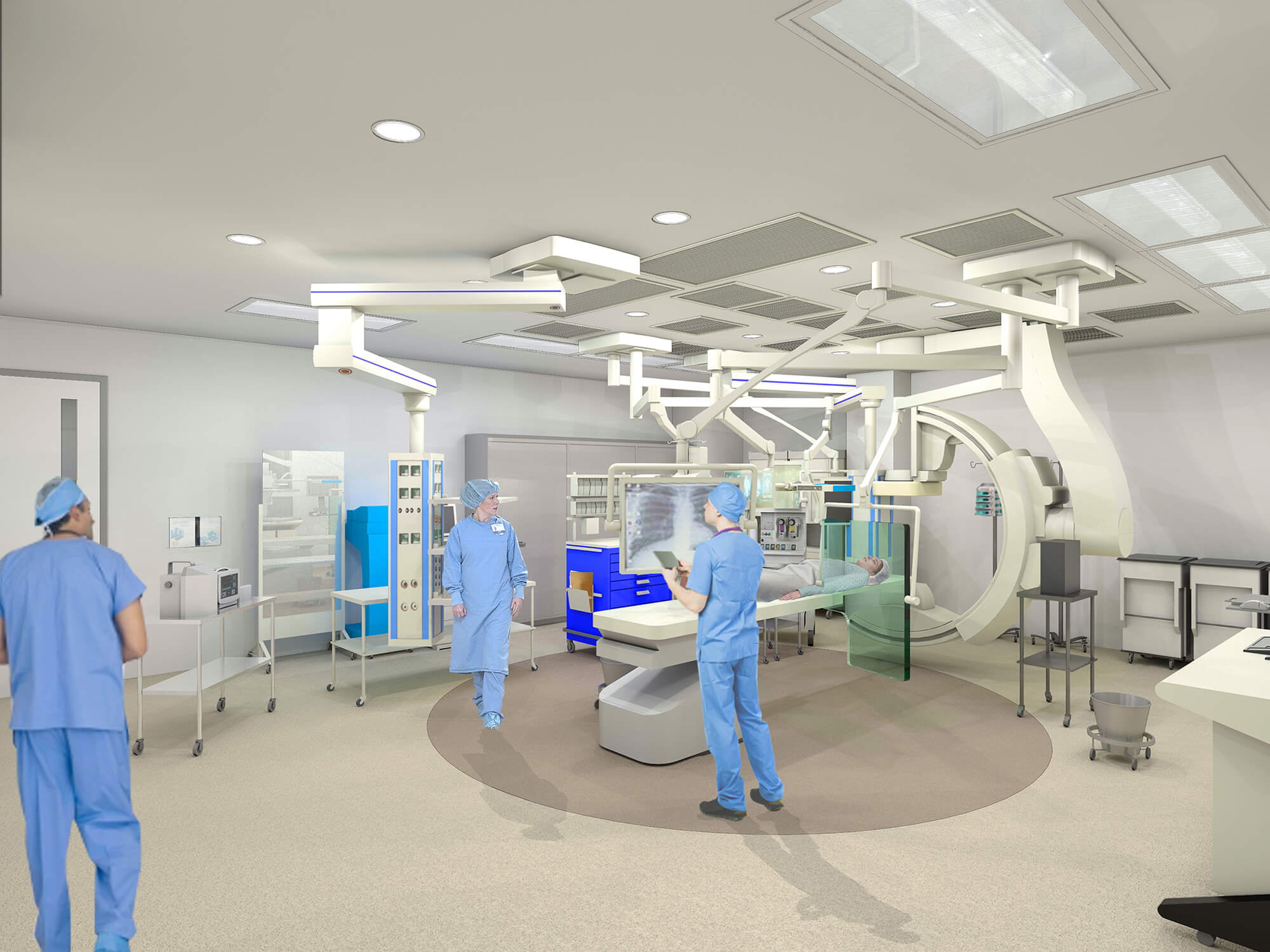
An electrophysiology (EP) study is a test used to evaluate and map the heart’s electrical activity and to diagnose arrhythmias. During an EP study, the electrophysiologist inserts thin wire electrodes into a vein in the neck or groin and then uses fluoroscopic imaging to thread the electrodes up to the heart. The EP study is often used to diagnose an arrhythmia, and allow for ablation as a treatment during the same procedure.
Ablation or Catheter Ablation
Abnormal electrical pathways in the heart that disrupt the cardiac electrical signal cause certain types of rapid or irregular heartbeats, known as tachycardias or arrhythmias. An ablation procedure is a treatment that uses a specialized catheter placed inside the heart. The catheter is placed via a needle poke in the leg vein, and threaded into the heart. It is capable of delivering energy to destroy the abnormal pathways, restoring the normal heart rhythm.
Our state-of-the-art EP lab allows us to offer the latest ablation technologies, including both cryoablation and radiofrequency ablation. In some patients, ablation can be used to treat common conditions such as supraventricular tachycardia (SVT), atrial fibrillation (AF), and premature ventricular complexes (PVCs).
Our center is a regional referral center for ablation of complex and life-threatening arrhythmias such as ventricular tachycardia (VT), including VT in a structurally compromised heart. MarinHealth offers the full complement of techniques for management of this condition. We use epicardial and endocardial approaches, and can insert temporary heart pumps to support the blood pressure during mapping. We have the latest tools for arrhythmia mapping and imaging to make these procedures safe and effective.
Cardioversion
Usually performed as a treatment for AFib or atrial flutter that does not stop on its own (a “persistent” pattern of these arrhythmias), cardioversion is a procedure to at least temporarily convert the heart’s rhythm back to normal. Depending on the circumstances, cardioversion can be used in combination with medications or procedures to control persistent atrial fibrillation or flutter. The procedure uses a short-acting deep sedative to allow a mild electric shock to convert the heart into a normal rhythm. Patients are discharged from this procedure same day (and often within an hour or so) with minimal recovery time needed. It is often performed in conjunction with a transesophageal echo to take pictures of the heart and exclude clot.
Pacemaker and Defibrillator Implantation
A pacemaker is a small, lightweight electronic device that’s implanted in the body to keep the heart from beating too slowly (bradycardia). The pacemaker keeps track of your heartbeat and, when necessary, generates electrical signals that keep your heart beating at the right pace. Newer pacemakers can transmit rhythm information to your doctor to provide remote monitoring of device function and rhythm health. They also can be programmed to adjust your heart rate in response to changes in your activity.
Our electrophysiology team can implant the full complement of implantable devices for treatment of rhythm disorders.
-
Single-chamber and Dual-chamber Pacemakers
These monitor and prompt one to two chambers of the heart to beat (generally the right atrium and right ventricle). This is accomplished via leads placed in the heart chamber(s) and attached to the generator box of the pacemaker.
-
Rate-adaptive Pacemakers
Single- or dual-chambered, these devices change the rate of the heartbeat in cases where the heart rate does not speed up naturally with physical activity.
-
Biventricular Pacemakers
These are used for cardiac resynchronization therapy. Leads in both the right and the left ventricle allow the pacemaker to literally resynchronize the action of the two chambers. This often results in notable improvement of cardiac function and a reduction in the severity of symptoms.
-
Leadless Pacemaker
Unlike traditional pacemakers, leadless devices such as the AVEIR™ DR Dual Chamber Leadless Pacemaker System are implanted directly into the heart through a minimally invasive procedure and eliminate the need for cardiac leads. As a result, leadless pacemakers reduce people’s exposure to potential lead and pocket-related complications and offer a less restrictive and shorter recovery period post-implantation.
-
Implantable Cardioverter Defibrillator Devices
These devices are implanted in a similar fashion as pacemakers; however, they are used to monitor for certain potentially lethal arrhythmias, and provide prompt, up-front pacing and/or shock therapy to restore a normal rhythm. They can be lifesaving in appropriate patients, particularly those with a history of compromised heart function, heritable arrhythmia disorders, or history of cardiac arrest. We offer both transvenous defibrillator device implantations (which are performed much like pacemaker implantations) and entirely subcutaneous (under the skin) defibrillator implants.
Ambulatory External Electrocardiogram (ECG ) Monitoring
Cardiac arrhythmias may occur infrequently and last for a short amount of time. Some arrhythmias occur only during exertion. By the time a person seeks help, the arrhythmia may have stopped, making a diagnosis impossible. Ambulatory ECG monitoring is used to record heart rhythm over days or even weeks in order to capture and record brief, intermittent arrhythmias.
Implantable Loop Recorder
Inserted just beneath the skin of the chest, this heart-monitoring device can record your heart rhythm for up to three years. Each night while you sleep, a record of your heart’s electrical impulses is sent to your doctor via a transmission monitor you keep beside your bed. A variety of patient-wearable devices, monitors, and implantable devices are now available to us. Even for the most well-informed patient, the selection can be difficult to navigate. Our experts are happy to consult with you about whether your situation is best managed through the use of a device, and if so, which one would be best for you.


.jpg)
.jpg)
-west-wing_Option-1.jpg)
-south-wing_Option-1.jpg)


/
View All
Electrophysiology Procedures Practitioners
Electrophysiology Procedures Locations
-
 MarinHealth Cardiovascular Performance Center | A UCSF Health Clinic
75 Rowland Way
More Information
Suite 250
Novato, CA 94945
415-927-0666 -
MarinHealth Cardiovascular Medicine | A UCSF Health Clinic
2 Bon Air Road
More Information
Suite 100
Larkspur, CA 94939
415-927-0666 -
MarinHealth Cardiovascular Medicine | A UCSF Health Clinic
75 Rowland Way
More Information
Suite 250
Novato, CA 94945
415-878-2910 -
MarinHealth Cardiovascular Medicine | A UCSF Health Clinic
651 First Street West
More Information
Suite L
Sonoma, CA 95476
707-935-1470