
Electrophysiology Conditions Treated
Atrial Fibrillation (AFib)
As your heart beats, the top chambers of the heart, or atria, contract
and push blood into the bottom chambers, or ventricles. In atrial fibrillation,
coordination with the ventricles is disrupted due to electrical abnormalities
in the pulmonary veins of the left atrium. This causes the atria to beat
chaotically and irregularly. Symptoms may include heart palpitations,
weakness, and shortness of breath. Atrial fibrillation is the most common
arrhythmia.
Stroke Risk from AFib
In AFib, there is a tendency for blood clots to form in the atria. 90%
of these clots form in a small pouch of the left atrium called the left
atrial appendage. If these clots come loose and travel to the brain, they
can block blood flow and cause a stroke. Stroke risk from AFib can be
reduced through the use of anticoagulant medication (blood thinners).
For people who live an active lifestyle, and/or do not tolerate blood
thinners well, the
WATCHMAN™ implant is a long-term solution for reducing stroke risk in AFib patients.
Atrial Flutter
Atrial flutter originates from an abnormal electrical impulse in one of
the atria. The atria beat regularly but faster and more frequently than
the ventricles, resulting in as many as four atrial beats to one ventricular
beat. Like AFib, atrial flutter is a condition that can increases the
risk of stroke due to the potential formation of blood clots in the heart.
Heart Block
The atrioventricular node (AV node) is a cluster of specialized cells between
the atria and the ventricles of the heart. The job of the AV node is to
channel electrical impulses from the atria to the ventricles. Heart block
is a conduction disorder in which the electrical signal from the AV node
to the ventricles is partly or completely blocked, resulting in an abnormally
slow heartbeat. Heart block is rated as first, second, or third degree,
with third degree being complete failure of electrical conduction.
Paroxysmal Supraventricular Tachycardia (PSVT)
Paroxysmal supraventricular tachycardia, or PSVT, is a sudden abnormal
rapid heartbeat that can come and go. The rapid heartbeat may last for
a few minutes or continue for hours. PSVT may have a variety of causes.
It doesn’t necessarily require treatment but it can be a symptom
of a variety of conditions so it’s a good idea to have it checked out.
Sinus Node Dysfunction
The timing of your heartbeat is regulated by a structure in the atria called
the sinus node–the heart’s natural pacemaker. Damage to the
sinus node, whether from surgery, drugs, a congenital heart defect, or
other causes, results in sinus node dysfunction, or “sick sinus
syndrome.” This may cause the heart rate to be irregular, too rapid
(tachycardia), or too slow (bradycardia).
Supraventricular Tachycardia (SVT)
Supraventricular tachycardia (SVT) is a general term for an abnormally
fast heart rhythm caused by faulty electrical activity in the upper part
of the heart. Atrial fibrillation (AFib), atrial flutter, paroxysmal supraventricular
tachycardia (PSVT), and Wolff–Parkinson–White syndrome are
all different types of SVT.
Vasovagal Syncope
Commonly known as a fainting spell, vasovagal syncope is caused by a sudden
drop in heart rate and blood pressure. Many people can faint in reaction
to a shocking or fearful situation. Common triggers include stress, heat
exposure, long periods of standing, and the sight of blood. While vasovagal
syncope is often a benign condition, in rare recurrent, persistent cases,
a pacemaker may be necessary.
Ventricular Tachycardia
As its name suggests, this abnormally rapid heart rhythm is caused by abnormal
electric signals in the ventricles of the heart. Ventricular tachycardia
may have several causes, including structural heart disease. It can occur
both in people with normal hearts and in people with certain types of
structural heart disease. It is often treated with implantable defibrillators.
Wolff-Parkinson-White (WPW)
In normal hearts, the AV node is the only connection between the upper
and lower chambers of the heart. The electrical signal that triggers a
heartbeat passes from the atria, through the AV node, and onto the ventricles.
People with Wolff-Parkinson-White syndrome are born with an extra muscle
fiber connecting the atria to the ventricles, creating an abnormal extra
electrical connection that can trigger a dangerously rapid heartbeat.
Wolff-Parkinson-White syndrome can lead to sudden cardiac death, a risk
that can be minimized with catheter ablation.

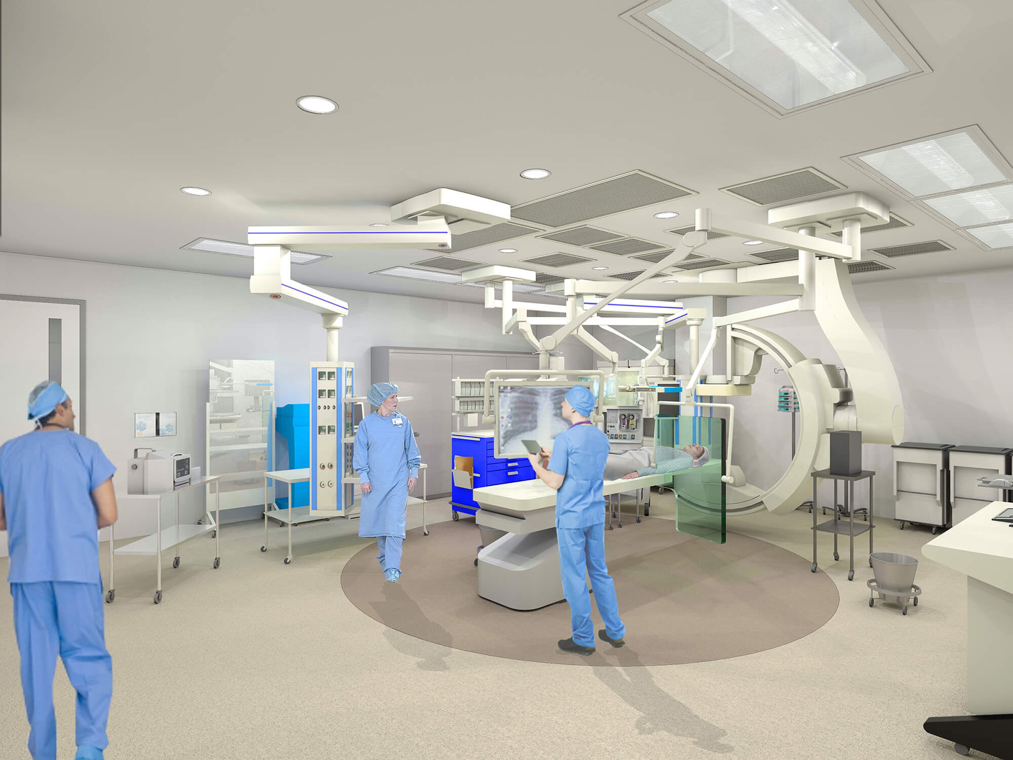
.jpg)
.jpg)
-west-wing_Option-1.jpg)
-south-wing_Option-1.jpg)


/
View All
Electrophysiology Conditions Practitioners
Electrophysiology Conditions Locations
-
 MarinHealth Cardiovascular Performance Center | A UCSF Health Clinic
75 Rowland Way
More Information
Suite 250
Novato, CA 94945
415-927-0666 -
MarinHealth Cardiovascular Medicine | A UCSF Health Clinic
2 Bon Air Road
More Information
Suite 100
Larkspur, CA 94939
415-927-0666 -
MarinHealth Cardiovascular Medicine | A UCSF Health Clinic
75 Rowland Way
More Information
Suite 250
Novato, CA 94945
415-878-2910 -
MarinHealth Cardiovascular Medicine | A UCSF Health Clinic
651 First Street West
More Information
Suite L
Sonoma, CA 95476
707-935-1470